|
Special X-Ray Procedures
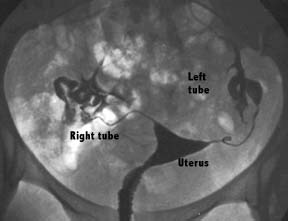
HYSTEROSALPINGOGRAM (HSG)
The procedure is performed to determine the size, shape, and position of the uterus and uterine tubes; to delineate lesions such as polyps, submucus tumor masses, or fistulous tracks; and to investigate the patency of the uterine tubes in patients who have been unable to conceive.
Contrast is injected via a catheter or cannula into the uterus to outline the uterine cavity and to delineate the fallopian tubes.
A normal study will show contrast spilling out of the tubes into the pelvis. The test must be performed within ten days of the onset of menstruation to ensure there is no chance of pregnancy.
The patient is placed in a standard gynae position on the edge of the screening table. The entrance to the vagina is cleansed with an antibacterial solution.
A vaginal speculum is inserted to obtain a view of the cervix. A small gauge rubber catheter or a cannula is inserted into the cervical canal. After repositioning, contrast is injected and several films obtained.
Following the procedure some discharge and minimal bleeding may occur. Although uncommon, increasing discharge, pain or fever could indicate a developing infection requiring treatment with antibiotics.
Preparation:
There is no preparation required.
WHAT TO EXPECT DURING A HYSTEROSALPINGOGRAM:
The woman lies on the table on her back and brings her feet up into a "frog leg" position.
The doctor places a speculum in the vagina and visualizes the cervix.
Either a soft, thin catheter is placed through the cervical opening into the uterine cavity or an instrument called a tenaculum is placed on the cervix and then a narrow metal cannula is inserted through the cervical opening.
Contrast is slowly injected through the cannula or catheter into the uterine cavity. An x-ray picture is taken as the uterine cavity is filling and then additional contrast is injected so that the tubes should fill and begin to spill into the abdominal cavity. Additional x-rays are taken as this "fill and spill" occurs.
When both tubes are demonstrated to be patent (or blocked), the woman is usually asked to roll to one side or the other slightly to give a slightly oblique x-ray image, which may help to further delineate her anatomy.
The procedure is now complete. The instruments are removed from the cervix and vagina. The woman usually remains on the table for several minutes to recover from the cramping which usually accompanies injection of the contrast.
After several minutes, the woman can get dressed and leave the centre.
The results of the test are immediately available. The x-ray pictures can be reviewed with the woman several minutes after the procedure has been completed if both she and the physician prefer to do this.
Pregnancy rates in several studies have been reported to be slightly increased in the first months following a hysterosalpingogram. This may be due to the fact that the flushing of the tubes with the contrast could open a minor blockage or clean out some debris that may be a factor that is preventing the couple from conceiving. Some of these studies suggest that using oil-based contrast provides a greater increase in pregnancy rates after a hysterosalpingogram than does the use of water based contrast.
INTRAVENOUS PYELOGRAM (IVP)
An IVP is an examination of the kidneys, ureters and bladder. An injection of contrast is given via an arm vein. The kidney rapidly excretes the contrast.
A series of films are obtained in sequence to follow the contrast from the kidneys, down the ureters and into the bladder. About five exposures in all are obtained in a routine series.
A tight compression band is often used across the middle of the abdomen during the procedure to better distend the upper ureters and kidney collecting systems. The technique may be modified in patients with acute pain (renal colic) or for follow up studies.
Preparation:
Bowel prep is required including a light low fiber diet, a period of fasting and laxatives.
Medications should be continued. A history of any previous allergy or asthma is obtained.
Note that no preparation is required for urgent cases.
URETHROGRAM
In a urethrogram, a small rubber catheter is inserted into the penis under aseptic conditions. Contrast is injected into the penis to delineate the penile urethra to look for narrowing, leakage of contrast indicating a rupture or abnormal passages.
Preparation: There is no preparation required.
VENOGRAM
A venogram requires an injection of contrast into a vein usually in the arm or foot with the aim of delineating the anatomy of the veins.
Tourniquets are used to slow the rate of flow of contrast in the veins and to direct flow into the deeper veins.
The technique was used frequently to look for blood clots in the leg veins and to display incompetent or varicose veins.
This information is now more easily obtained using Vascular Ultrasound.
Preparation:
A history of any previous allergy or asthma is obtained.
|